Anatomy Of Upper Thigh And Hip - Hip Anatomy / Iliopsoas muscle, a hip flexor muscle that attaches to the upper thigh bone.. He also serves the communities of charleston, sc and augusta, ga. The hip's unique anatomy enables it to be both extremely strong and amazingly flexible, so it can bear weight and allow for a wide range of movement. Iliopsoas muscle, a hip flexor muscle that attaches to the upper thigh bone. Diarthrodial joint with its inherent stability dictated primarily by its osseous components/articulations. Hip surgeon dr guillaume dumont offers hip pain treatments in columbia, sc.
The adductor muscle on the inner thigh; Tibial part of the sciatic nerve action: The anatomical areas found on the upper limb can serve as key landmarks to help us find important anatomical structures such as finding one of the superficial veins: It functions to adduct the thigh and to flex. During hip replacement surgery, your surgeon removes the upper part of your thigh bone, including the femoral head (ball of the hip joint) and a part the upper part of the thigh bone is then exposed, and a series of tools called broaches are introduced one at a time to prepare your thigh bone for a metal.
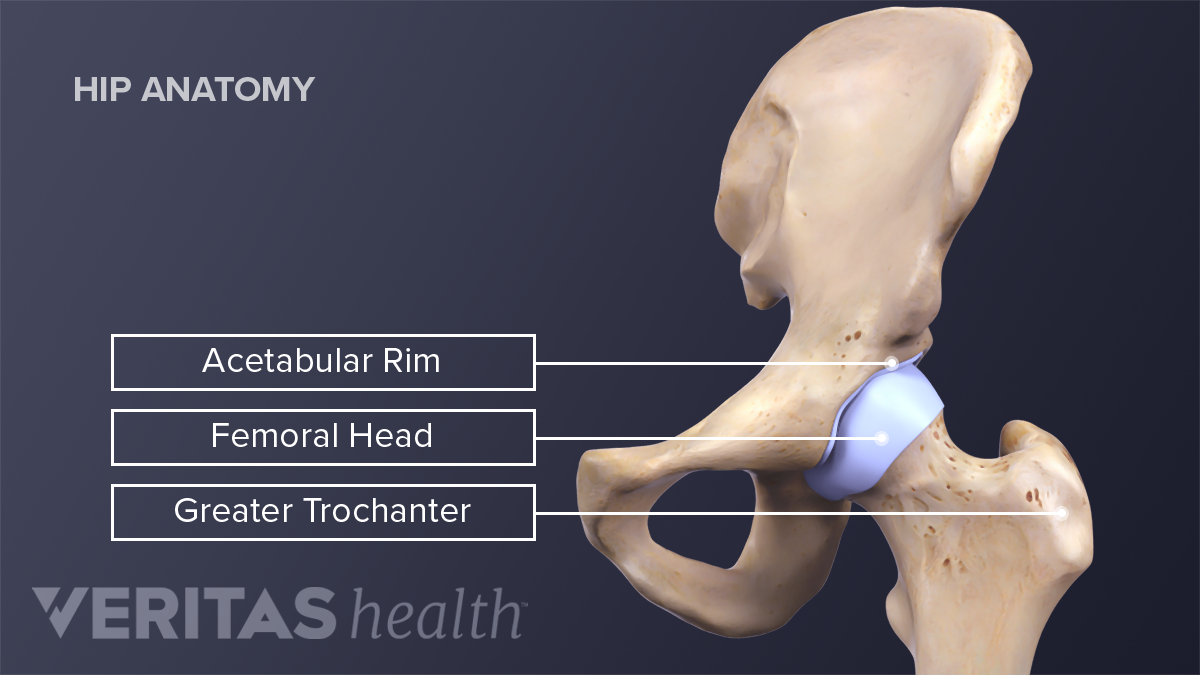
3d Human Upper Leg Anatomy Or Anatomical And Muscle Set Stock Illustration Illustration Of Foot Health 198223856 from thumbs.dreamstime.com Foundational anatomy provides medical students with the necessary background in anatomy for success in clerkships. This webpage presents the anatomical structures found on thigh mri. 431).—at the upper and medial part of the thigh, a little below the medial end of the inguinal ligament, is a large. The upper part of the thigh bone consists of the femoral head, femoral neck, and greater and lesser trochanters. Several muscles cross the front of the hip and create hip flexion, pulling the thigh and trunk toward both muscles cross the floor of the pelvis, emerge at the outer edges of the pubic bones, and finally insert on the inner upper femur (thighbone). The adductor muscle on the inner thigh; May 13, 2019 edited by dr. Iliopsoas muscle, a hip flexor muscle that attaches to the upper thigh bone.
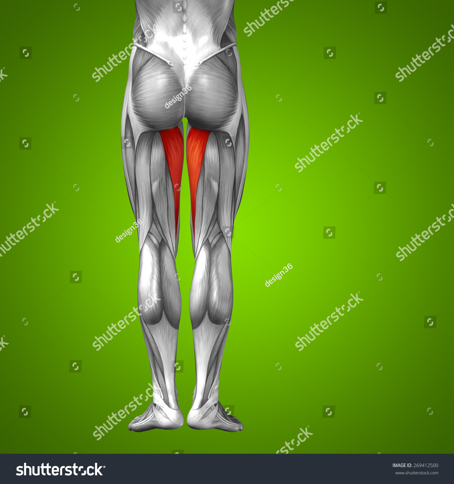
Chief flexor of knee weak.
The following nerves serve the gluteal and. for detailed anatomy of pelvic bones, read anatomy of hip bone. On the anterior side, the most prominent of the muscles are the sartorius muscle and the four the four muscle of the quadriceps all extend the lower leg, and the rectus femoris additionally can flex the thigh at the hip. Hip surgeon dr guillaume dumont offers hip pain treatments in columbia, sc. Use the mouse scroll wheel to move the images up and down alternatively use the tiny arrows (>>) on both side of the image to move the images. This deep muscle begins in the low back and pelvis and connects on the inside edge of the upper femur. The femur, the hip bone (subdivided into ilium. May 13, 2019 edited by dr. Want to learn more about it? Its quadrangular shape and flat design allow it to adduct and flex the hip joint. Hip and knee pain and hip and shoulder pain are. The hip joint is a ball and socket joint that is the point of articulation between the head of the femur and the acetabulum of the pelvis. This webpage presents the anatomical structures found on hip mri.
Chief flexor of knee weak. The hip's unique anatomy enables it to be both extremely strong and amazingly flexible, so it can bear weight and allow for a wide range of movement. 431).—at the upper and medial part of the thigh, a little below the medial end of the inguinal ligament, is a large. Hip anatomy, function and common problems. The anatomical areas found on the upper limb can serve as key landmarks to help us find important anatomical structures such as finding one of the superficial veins:
Hip Anatomy from embed.widencdn.net This arrangement gives the hip anatomy a large amount of motion needed for daily activities. This deep muscle begins in the low back and pelvis and connects on the inside edge of the upper femur. Bones of the lower limb. It functions to adduct the thigh and to flex. Knowing the anatomy of your hip can help you understand the source of any hip pain. 340 anatomical structures of the hip region were labeled, accessible on anatomical parts: While the thigh muscles will be slip into the anterior, medial and posterior groups. The anatomical areas found on the upper limb can serve as key landmarks to help us find important anatomical structures such as finding one of the superficial veins:
B, muscles of the anterior thigh compartment.
Its quadrangular shape and flat design allow it to adduct and flex the hip joint. The following nerves serve the gluteal and. Atlas of human anatomy in cross section. This vein, as well as the deep veins. The joints and muscles of the hips and thighs need nervous input so they can do what your brain wants them to do. Use the mouse scroll wheel to move the images up and down alternatively use the tiny arrows (>>) on both side of the image to move the images. It's restricted by contact of the thigh with all the abdomen and adduction is restricted by contact. This mri hip joint axial cross sectional anatomy tool is absolutely free to use. 431).—at the upper and medial part of the thigh, a little below the medial end of the inguinal ligament, is a large. Diarthrodial joint with its inherent stability dictated primarily by its osseous components/articulations. Groin, inguinal region and the anterior and posterior regions of the hip and thigh. Iliopsoas muscle, a hip flexor muscle that attaches to the upper thigh bone. The upper part of the thigh bone consists of the femoral head, femoral.
Unlike the shoulder girdle, the pelvic girdle is firmly integrated into the axial skeleton: 431).—at the upper and medial part of the thigh, a little below the medial end of the inguinal ligament, is a large. All of the anatomical parts of the hip work together to enable various movements. In vertebrate anatomy, hip (or coxa in medical terminology) refers to either an anatomical region or a joint. Anatomy ▶ lower limb ▶ bones and cartilages ▶ hip joint.
Concept Conceptual 3d Human Upper Leg Stock Illustration 269412500 from image.shutterstock.com Hip flexor deep in pelvis a composite o… used to extend the hip when climbing st… The thigh is the area between the hip and the knee joint. The upper part of the thigh bone consists of the femoral head, femoral neck, and greater and lesser trochanters. Unlike the shoulder girdle, the pelvic girdle is firmly integrated into the axial skeleton: The femur, the hip bone (subdivided into ilium. Groin, inguinal region and the anterior and posterior regions of the hip and thigh. In vertebrate anatomy, hip (or coxa in medical terminology) refers to either an anatomical region or a joint. It functions to adduct the thigh and to flex.
Hip movements include flexion, extension, abduction, adduction, circumduction, and hip rotation.
Hip and leg pain can cause stress on joints and affect other areas of the body. This vein, as well as the deep veins. Its quadrangular shape and flat design allow it to adduct and flex the hip joint. Pelvis, perineum, hip, and upper thigh. The muscles also require a lot of blood flow, which provides oxygen and nourishment, especially when you're physically active. The paired hip bones are connected. The femur or thigh bone is one of the longest bones in the human body. Anatomy ▶ lower limb ▶ bones and cartilages ▶ hip joint. The iliopsoas muscle, which extends from the lower back to upper femur; All of the anatomical parts of the hip work together to enable various movements. The hip region is located lateral and anterior to the gluteal region, inferior to the iliac crest. On the anterior side, the most prominent of the muscles are the sartorius muscle and the four the four muscle of the quadriceps all extend the lower leg, and the rectus femoris additionally can flex the thigh at the hip. Anatomy hip, thigh and leg muscles.
Groin, inguinal region and the anterior and posterior regions of the hip and thigh upper thigh anatomy. Bones of the lower limb.


0 Komentar